CBCT: When a 3D Scan Is Justified
A cone-beam CT scan can reveal anatomy hidden by a flat dental X-ray, but it also exposes the patient to more radiation. The right question is not whether 3D imaging is impressive. It is whether the additional information will change diagnosis, treatment, or safety.
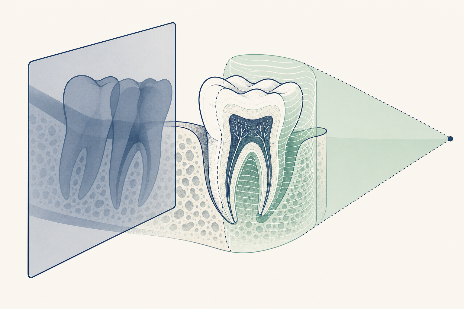
The dose question starts with the clinical question
A conventional intraoral X-ray compresses three-dimensional anatomy into a two-dimensional image. Structures can overlap, root canals can be hidden behind one another, and the thickness of bone cannot be measured reliably from a flat image. CBCT works differently. A rotating cone-shaped X-ray beam records multiple projections, and software reconstructs them into axial, coronal, sagittal, and three-dimensional views. That extra information is not free. Dental CBCT generally delivers more radiation than an intraoral, bitewing, or panoramic radiograph, although it is usually lower dose than a medical CT examination of a comparable region. The relevant principle is justification: the scan should answer a defined clinical question, and the expected benefit should outweigh the radiation risk. A scan ordered because the machine is available, because every new patient receives one, or because a 3D image looks more advanced is not a sound indication. For a patient, the practical test is simple: what decision will this scan change? If the answer is unclear, a lower-dose two-dimensional image or no image may be more appropriate.
A 3D scan is justified by the decision it enables, not by the fact that it produces a 3D picture.
What changes in the chair
The scan itself is usually quick and non-invasive. The patient stands or sits while the scanner rotates around the head. The teeth may be separated with a small bite block, and the head must remain still. There is no injection and usually no preparation. The machine produces a volume of data rather than a single film, allowing the clinician to scroll through thin slices and inspect anatomy from different directions. The patient may notice little difference during the scan, but the treatment conversation can change substantially. An implant plan can account for the mandibular canal, mental foramen, maxillary sinus, nasal floor, and the width and contour of the alveolar ridge. An endodontic assessment can reveal an extra canal, a missed root, a perforation, a fractured root, or a lesion that is difficult to localise on periapical films. Surgical planning can show the relationship between an impacted tooth and the inferior alveolar nerve or adjacent tooth roots. CBCT does not automatically produce a diagnosis. The volume must be selected, acquired, reconstructed, and interpreted correctly. A limited field of view is often preferable when the question concerns one tooth or a small surgical site. A larger scan should not be used simply because it is easier to obtain.
The patient feels almost nothing during CBCT. The value lies in the anatomy that becomes measurable afterward.
Implant planning is one of the clearest indications
Before implant placement, the clinician needs more than a rough view of the missing tooth. The proposed implant must fit within the available bone while avoiding vital structures and supporting the planned crown. CBCT can show the buccolingual width of the alveolar ridge, the height beneath the maxillary sinus, the position of the mandibular canal, and concavities on the lingual or facial surface that may be invisible on a panoramic image. This information can affect the implant diameter and length, the angulation, the need for bone augmentation, the timing of treatment, and whether an alternative prosthetic plan is safer. In the posterior maxilla, the sinus floor and residual ridge can be assessed in cross-section. In the posterior mandible, the inferior alveolar canal can be traced before surgery. In the anterior maxilla, the facial plate and incisive canal may influence implant position and soft-tissue support. CBCT should be integrated with a clinical examination, periodontal assessment, diagnostic casts or an intraoral scan, and the planned restoration. Digital implant planning may merge CBCT data with a surface scan so the implant is positioned for the final crown, not merely placed where bone happens to be present. The scan supports planning, but it does not remove the need for surgical judgment.
For implants, CBCT turns hidden bone thickness and nearby structures into planning information.
Endodontics: when a flat film leaves competing explanations
Root canal diagnosis often begins with history, clinical tests, and carefully angled periapical radiographs. CBCT is not a routine screening test for every painful tooth. It becomes more defensible when symptoms, examination, and two-dimensional images do not agree, or when the result is likely to alter treatment. A limited-volume scan may help identify untreated canals, complex root morphology, apical pathology, root resorption, perforation, separated instruments, or the relationship of a lesion to the cortical plates and neighbouring roots. For example, a maxillary first molar may have a second mesiobuccal canal that is difficult to detect in a single projection. A mandibular premolar may have unusual canal anatomy, while a persistent lesion after root canal treatment may require distinction between missed anatomy, fracture, perforation, or non-endodontic disease. The scan must be interpreted with awareness of artefacts from crowns, posts, amalgam, and other dense materials. Beam hardening can create dark bands or streaks that mimic defects. CBCT also has limited ability to prove a vertical root fracture in every case. The correct sequence is clinical assessment first, targeted radiographs where useful, and CBCT when the unresolved question matters to the treatment choice.
CBCT is most useful in endodontics when it resolves a specific disagreement between symptoms, tests, and flat images.
Surgery, impacted teeth, and anatomy that overlaps
A panoramic image can suggest where an impacted mandibular third molar lies, but overlapping roots and the mandibular canal may leave the surgical relationship uncertain. CBCT can show whether the canal passes buccal, lingual, or between the roots, whether the roots are curved or fused, and whether there is cortical plate thinning. That may influence the choice between removal, monitoring, coronectomy, or referral, although the scan does not predict every surgical outcome. The same principle applies to impacted canines. CBCT can locate the tooth in three dimensions, assess root resorption on adjacent incisors, and help the orthodontic and surgical team plan exposure and traction. It can also assist in evaluating jaw lesions, facial asymmetry, cleft-related anatomy, and selected trauma cases when a two-dimensional image cannot define the fracture or displacement. For temporomandibular joint assessment, CBCT is primarily a hard-tissue examination. It can demonstrate condylar shape, cortical changes, erosions, osteophytes, and gross bony asymmetry. It does not show the articular disc as well as magnetic resonance imaging, and it should not be presented as a universal explanation for facial pain or joint symptoms. The scan should cover the smallest region that answers the question, and all relevant anatomy within that volume must be reviewed.
CBCT earns its place when overlap conceals a nerve, root, fracture, lesion, or surgical boundary.
Where the marketing runs ahead of the evidence
A CBCT image can make a consultation feel more authoritative without making the underlying decision more accurate. Three-dimensional visuals are particularly persuasive in implant, orthodontic, airway, and cosmetic treatment marketing. That visual confidence should not be confused with clinical necessity. CBCT is not a general health scan, a routine cavity screen, or a substitute for examination. It does not prove that a patient needs an implant, orthodontic treatment, airway surgery, or treatment for temporomandibular symptoms. It cannot by itself establish whether a tooth is restorable, whether pain is odontogenic, or whether a radiolucent area is benign or malignant. Findings may need comparison with previous images, clinical tests, specialist review, or another modality. Airway measurements deserve particular caution. A CBCT can display the airway at one moment and in one head position, but the image is not automatically a diagnosis of obstructive sleep apnoea. The scan should not be sold as a screening test for every snoring patient. Similarly, artificial intelligence may help with segmentation, measurements, or image triage, but software output remains dependent on image quality, training data, and human verification. The proven benefit is question-specific anatomy. The marketing claim is that more data always means better care. It does not.
More anatomy is not the same as more diagnosis.
A defensible scan has a disciplined workflow
A sound CBCT pathway begins with the record: symptoms, examination, existing radiographs, medical and imaging history, and the decision that remains unresolved. The operator then chooses the smallest field of view and the lowest exposure setting that can provide adequate diagnostic quality for the patient’s size and the indication. Low dose should not mean unusable images, and high resolution should not be selected without a reason. Before exposure, the clinician should confirm the correct patient, region, and purpose, remove avoidable artefacts where appropriate, and position the patient carefully. Movement can blur the volume and lead to a repeat scan. After acquisition, the clinician must review the entire volume within the field of view, not only the tooth or implant site that prompted the referral. Significant incidental findings should be documented and referred appropriately. Interpretation should be performed by a practitioner with suitable training, with specialist reporting when the findings or scope require it. The patient should receive a plain explanation: why the scan is needed, what lower-dose alternatives were considered, what area will be scanned, and how the result may change treatment. Pregnancy status and previous relevant imaging should be discussed according to local clinical policy. The scan should be stored and shared in a way that avoids unnecessary duplication.
Justification, optimisation, accurate interpretation, and follow-up are one process, not separate technical details.
The answer for patients and practices
For a patient, the right question is not whether CBCT is safe in the abstract. It is whether this particular scan is necessary for this particular decision. Ask what the clinician is trying to see, whether an intraoral or panoramic X-ray would answer the question, whether the field of view can be limited, and who will interpret the complete volume. If a previous scan covers the same anatomy and remains clinically suitable, repeating it may add exposure without adding information. For clinicians and operators in the UAE and GCC, the same standard applies regardless of whether the scan is performed inside a dental clinic or at an imaging centre. Equipment availability and treatment-package design should not replace indication-based selection. Local licensing, quality assurance, operator training, record retention, and reporting requirements must also be followed. The strongest case for CBCT is a defined three-dimensional problem: implant site anatomy, a complex or unresolved endodontic case, an impacted tooth near a vital structure, selected trauma, or a lesion whose extent cannot be understood on two-dimensional imaging. The weakest case is routine scanning without symptoms, findings, or a treatment decision that depends on the result. CBCT is an important tool because it can expose anatomy that flat X-rays hide. It is responsible care only when that extra anatomy changes what happens next.
Use CBCT when three-dimensional information changes the plan. Do not use it to decorate a consultation.
Key findings
- CBCT provides cross-sectional information that conventional dental radiographs cannot reliably provide.
- The scan should be justified by a defined diagnostic or treatment question, with lower-dose alternatives considered first.
- Implant planning, selected complex endodontic cases, impacted teeth near vital structures, selected trauma, and assessment of some bony lesions are established areas of use.
- A limited field of view and patient-specific exposure settings can reduce unnecessary radiation while preserving useful image quality.
- CBCT does not replace clinical examination, conventional radiographs, specialist judgment, or other imaging modalities when they are more appropriate.
- Three-dimensional images can support planning but do not by themselves prove treatment need, predict outcomes, or diagnose airway disease.