Intraoral Scanners Hit a Tipping Point
Prices fell, the field consolidated, and the wand finally pays for itself. Our radar ranks scanners on cost, accuracy, and workflow lock-in, with a buy, wait, or skip verdict.
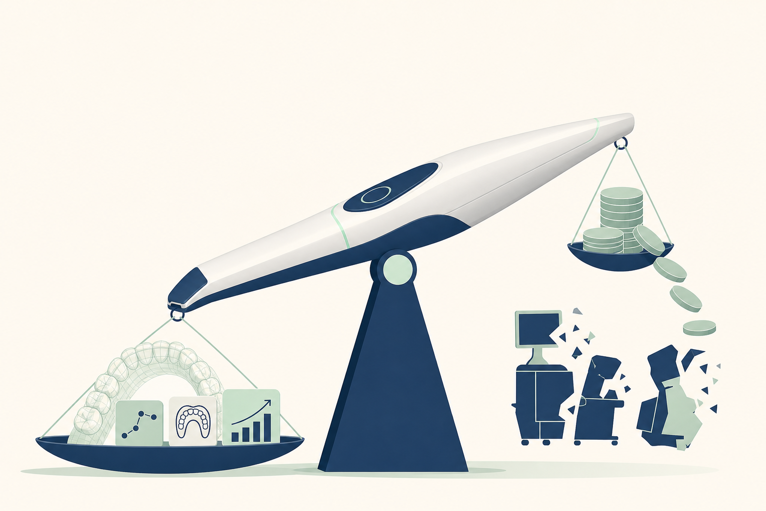
8-slide carousel · storyboard copy
The wand is changing dental visits
An intraoral scanner uses a small camera to capture the teeth, gums, bite, and nearby oral tissues as a digital model. No impression tray or setting alginate is required for many routine cases.
- Buy: practices using a scanner for restorations, aligners, retainers, or monitoring
- Wait: cases where the clinician needs to confirm the scanner fits the treatment workflow
- Skip: a scanner used only as a visual gadget without a clinical decision attached
Why it matters to patients
The scan records tooth surfaces, occlusal contacts, gingival margins, missing teeth, and the shape of the dental arches. The software stitches overlapping images into a three-dimensional file that can guide a laboratory or chairside CAD/CAM system.
- Accuracy depends on the scanner, operator, saliva control, scan path, reflective surfaces, and anatomy
- A dry field improves capture around the gingival sulcus and prepared tooth margins
- Long full-arch scans and completely edentulous mucosa can be more difficult than short scans of teeth
What happens during a scan
The clinician first examines the mouth, removes debris, controls saliva, and retracts the cheeks and tongue. The wand then records the occlusal, buccal, and palatal or lingual surfaces. The software aligns each new image with the previous one and flags gaps for rescanning.
- For a crown, the dentist prepares enamel and dentine, places a retraction cord or paste when needed, dries the finish line, and scans the preparation and opposing arch
- The patient bites together so the software can register the maxillary and mandibular arches
- The digital file may be sent for a ceramic, zirconia, lithium disilicate, resin, metal, aligner, or retainer design
The one rule that matters
A digital impression is only useful when the clinician checks it. The dentist should inspect the finish line, interproximal contacts, occlusion, soft-tissue capture, missing data, and the planned restoration before sending the file.
- A clean-looking 3D model does not prove that every margin or contact is clinically correct
- If blood, saliva, movement, reflective metal, or a deep subgingival margin hides anatomy, that area must be managed and rescanned
- The scanner supports diagnosis and fabrication; it does not replace examination, radiographs, or clinical judgement
Mythbuster: digital always means more accurate
Not automatically. For many tooth-supported restorations and orthodontic records, digital scans can provide a comfortable and clinically useful alternative to conventional impressions. Accuracy still changes with span length, implant position, edentulous tissue, preparation geometry, and scanning technique.
- Myth: the scanner can see through enamel or gum. Fact: it records visible surfaces and cannot replace radiographs for bone, roots, or hidden decay
- Myth: every scanner works with every laboratory. Fact: file formats, software subscriptions, export rules, and laboratory compatibility can create workflow lock-in
- Myth: faster scanning means better treatment. Fact: the final result depends on diagnosis, preparation, design, materials, milling or printing, and fit verification
Make the choice fit your treatment
Ask what the scan will change in your care. A scanner is most useful when it connects to a defined task such as a crown, bridge, implant restoration, aligner, night guard, denture record, or comparison of tooth wear and gum position over time.
- For a single crown, prioritise clear margin capture, bite registration, and laboratory compatibility
- For implants, confirm the scan body is seated, the implant platform is recorded, and the laboratory accepts the file and system
- For aligners or retainers, ask how the scan is reviewed, approved, and updated when teeth or attachments change
- For extensive tooth loss, ask whether conventional records or another digital method is more predictable for the planned prosthesis
When to see a dentist, not a sales demo
Book an examination if you have pain, swelling, bleeding that persists, a loose tooth, a broken restoration, sensitivity that does not settle, a change in your bite, or difficulty chewing. A scanner can document the problem, but treatment requires an examination and sometimes bitewing, periapical, panoramic, or three-dimensional imaging.
- Ask who owns the digital file and how it is protected
- Ask whether the scan is being used for a specific diagnosis or appliance
- Ask what happens if the scan is incomplete or the restoration does not fit
- A scan should not delay urgent care for facial swelling, fever, trauma, or uncontrolled bleeding
The verdict: useful tool, not magic
For patients, the best scanner is the one that produces a complete record inside a well-controlled clinical workflow. Buy when it improves a treatment you actually need. Wait when the practice cannot explain the workflow or file handling. Skip when it adds animation but no clinical purpose.
- Compare cost, scan completeness, laboratory compatibility, data ownership, and clinician experience
- The wand pays for itself only when it improves the care pathway, not when it is simply present in the room
- Save this carousel and ask your dentist which step of your treatment the scan will control