AI Diagnostics Crosses From Demo to Default
What caries and bone-loss AI can see, what it cannot decide, and whether it changes your treatment plan
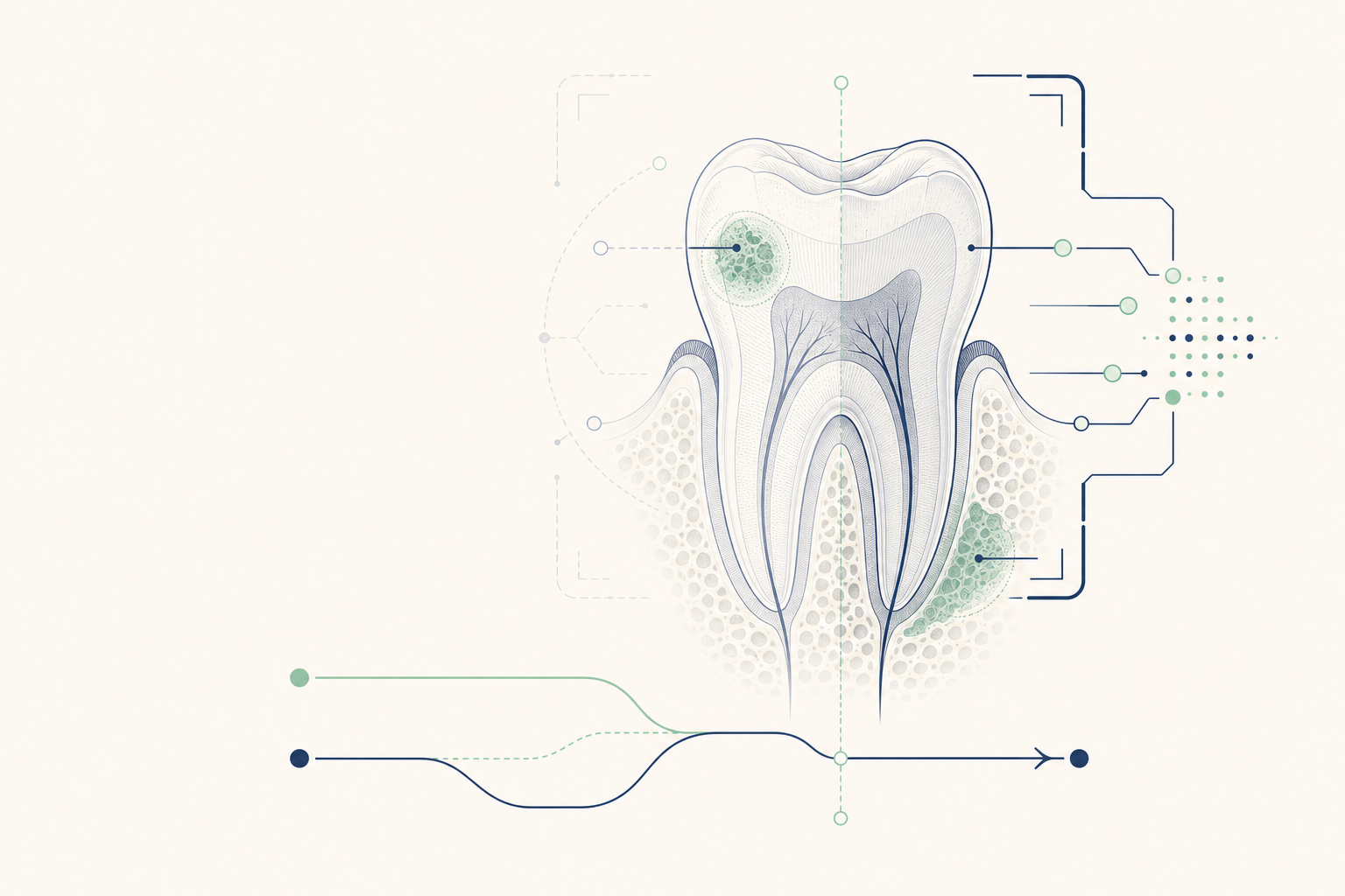
8-slide carousel · storyboard copy
AI is entering the operatory
Software can analyze dental X-rays and mark patterns linked with tooth decay and loss of supporting bone. It is an additional set of eyes, not an autonomous dentist.
- Used mainly with bitewing, periapical, and panoramic radiographs
- May display suspected lesions, boundaries, or bone levels
- The dentist still confirms the finding and explains its meaning
Why it matters to patients
Caries can begin beneath the contact between teeth or under an existing filling. Periodontal bone loss affects the alveolar bone around tooth roots and may progress before mobility or pain appears. AI can draw attention to areas that deserve a closer clinical review.
- A bitewing image may reveal enamel or dentine demineralisation between molars
- A panoramic or periapical image may show reduced crestal bone height
- AI cannot reliably determine activity, symptoms, bacterial risk, or the best treatment by itself
- Adoption is increasing, but rates differ between clinics and health systems
The protocol: image to decision
A safe workflow connects the image with the patient, not just the algorithm.
- 1. The dentist takes radiographs only when clinically justified and records the patient history and examination findings.
- 2. The software processes the digital image and may outline a radiolucent area, restoration margin, calculus, or distance from the cemento-enamel junction to the alveolar crest.
- 3. The dentist checks image quality, exposure, anatomy, restorations, overlap, artefacts, and the software's flagged area.
- 4. The dentist compares earlier images when available and examines the tooth, gingiva, periodontal pockets, bleeding, plaque, vitality, and symptoms.
- 5. The treatment plan changes only when the combined evidence supports it, such as monitoring an early non-cavitated lesion, repairing a defective restoration, or arranging periodontal therapy.
The one rule that matters
AI may suggest a finding. A qualified dentist must verify it before diagnosis or treatment.
- A bright mark is not automatically a cavity
- A lower bone level is not, by itself, proof of active periodontitis
- The dentist remains responsible for the diagnosis, consent, treatment, records, and follow-up
- Ask whether the finding is visible on the image, supported by the examination, and relevant to your treatment plan
Mythbuster: an AI flag is not a verdict
AI does not replace a clinical examination, and it does not guarantee that every disease is detected. False positives can lead to unnecessary concern or treatment. False negatives can occur when decay is hidden by overlapping anatomy, restorations, poor positioning, or image limitations.
- AI cannot measure bacterial activity inside a lesion
- AI cannot decide whether a tooth needs fluoride exposure, sealant, restoration, root canal treatment, or extraction
- AI cannot establish periodontal disease from an image alone
- A second review may be appropriate when the image and examination do not agree
What it means for your mouth
The same radiographic mark can lead to different actions in different patients. A small enamel lesion in a low-risk patient may be monitored with plaque control, fluoride toothpaste, diet advice, and a review. A cavitated dentine lesion that traps plaque may need a restoration. Bone loss with bleeding pockets and calculus may require periodontal examination and professional debridement.
- Your plan depends on lesion depth, cavitation, symptoms, restorability, caries risk, periodontal measurements, smoking, diabetes, medicines, and previous disease
- Interdental brushes are preferable where the space allows; floss remains useful where the contact is tight
- After brushing, spit out the toothpaste and do not rinse with water
- AI does not change the need for daily plaque removal or scheduled clinical reviews
Ask these questions at the appointment
Reimbursement and liability are not universal. Whether an AI-enabled review is covered depends on the payer, the coded service, the clinic, and the applicable UAE rules. A software flag does not transfer clinical responsibility away from the treating dentist.
- What image and anatomical finding did the software identify?
- Can you show me the finding and explain its depth or severity?
- What did the clinical examination and periodontal measurements add?
- Would the treatment plan be different without the AI flag?
- Is the AI review included in the visit, and how is it documented?
Better detection should mean better decisions
The useful question is not whether AI found something. It is whether verified information about your enamel, dentine, restorations, gingiva, periodontal pockets, and alveolar bone changes what should happen next.
- Request an explanation in plain language
- Keep previous radiographs available for comparison
- Proceed only after the diagnosis, alternatives, benefits, risks, and follow-up are clear
- Book a dental assessment if you have pain, swelling, bleeding, loose teeth, food trapping, or a broken filling