The Cost of Waiting
A delayed filling is not a neutral decision. As decay moves from enamel into dentine, the pulp, and eventually the tissues around the root, treatment becomes more invasive, less predictable, and harder to access. Dental Nation’s position is simple: transparent early care is both a clinical duty and a trust strategy for UAE clinics.
A small cavity is a time-sensitive problem
Dental caries begins when plaque bacteria metabolise fermentable carbohydrates and produce acids that lower the pH at the tooth surface. Enamel loses mineral first. At this stage, the lesion may be non-cavitated, appearing as a chalky white spot, and the surface can sometimes be managed with plaque control and topical fluoride rather than a restoration. Once the enamel surface collapses, bacteria and food can enter the cavity. The lesion then advances through enamel toward the dentinoenamel junction and into dentine. Dentine is softer and contains tubules that communicate with the pulp. A cavity in dentine can therefore enlarge laterally beneath apparently intact enamel. The patient may feel nothing because caries itself is not always painful. A clinician can identify the lesion through examination, bitewing radiographs where indicated, and assessment of plaque, diet, saliva, and previous disease. The first decision is not simply whether to drill. It is whether the lesion is active, cavitated, cleanable, and structurally capable of being sealed or restored. Dental Nation should argue that early diagnosis is the most honest form of cost communication. A patient cannot make a meaningful financial decision when the clinic explains only the price of the final procedure and not the biological process that makes delay more expensive.
The first cost of waiting is often invisible: mineral loss, bacterial access, and weakening tooth structure.
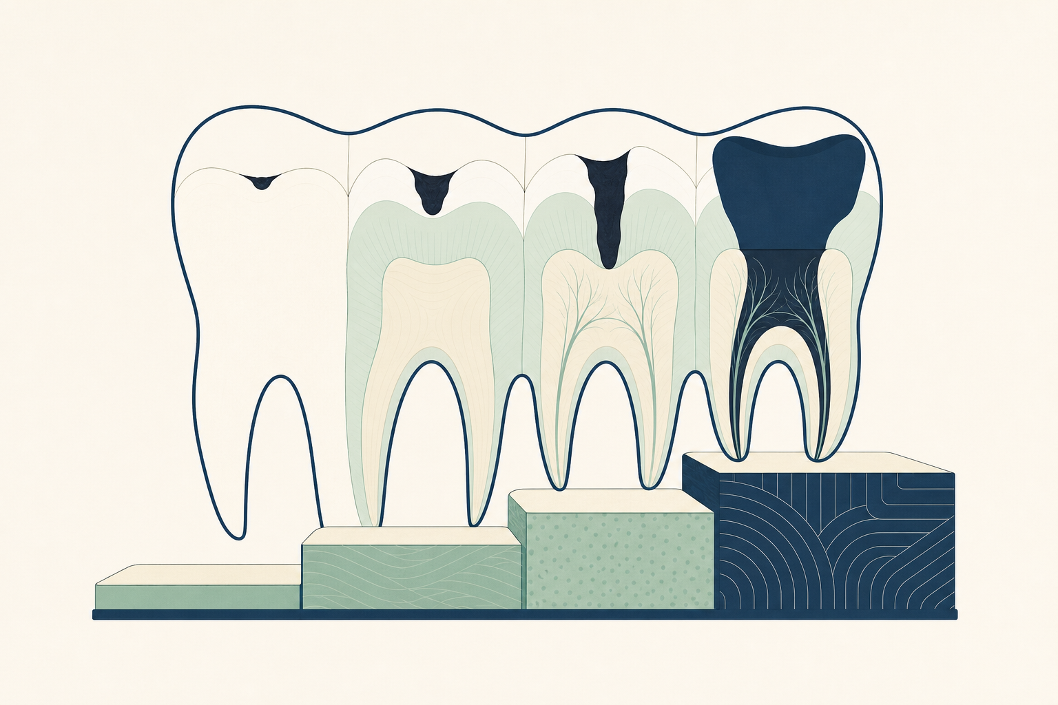
The treatment ladder follows the anatomy
The treatment ladder is not a sales ladder. It follows where the disease has travelled. A non-cavitated enamel lesion may receive preventive management, including improved brushing with fluoride toothpaste, interdental cleaning, dietary control, and professional fluoride application when appropriate. The patient should spit after brushing rather than rinse, because rinsing removes much of the concentrated fluoride left on the teeth. A small cavitated lesion that remains restorable may require removal of infected tooth tissue and placement of a direct restoration. The clinician isolates the tooth, removes diseased structure while preserving sound enamel and dentine, conditions the surface where required, and places a material such as resin composite or another indicated restorative material in increments. The restoration must reproduce the contact point, marginal seal, occlusal anatomy, and ability to clean around the tooth. The material choice depends on moisture control, cavity size, load, aesthetics, remaining walls, and caries risk. If decay has removed a substantial amount of tooth structure, a direct filling may no longer protect the cusps. An indirect restoration, such as an onlay or crown, may be indicated after assessment of remaining walls, cracks, occlusion, and periodontal health. If bacteria and inflammation have reached the pulp, restoration alone cannot resolve the disease. Root canal treatment, followed by a definitive coronal seal and sometimes cuspal coverage, becomes the relevant pathway.
The ladder rises because the disease changes the tooth, not because the clinic changes the product.
Pain is a poor timetable
Waiting for pain is a clinical error because pulp inflammation does not follow a patient-friendly schedule. Early dentinal decay may cause brief sensitivity to cold or sweet stimuli. As bacterial irritation approaches the pulp, the response can become prolonged, spontaneous, or difficult to localise. Reversible pulpitis may settle when the cause is removed and the tooth is sealed. Irreversible pulpitis may require pulpotomy or root canal treatment depending on the tooth, development, diagnosis, and treatment setting. A painful tooth is not always the most advanced tooth, and a painless tooth is not necessarily safe. The pulp can lose vitality while the patient experiences little warning. Necrotic pulp tissue can allow bacteria to move through the apical foramen into the periapical tissues. The result may be apical periodontitis, an acute abscess, swelling, tenderness to biting, or a draining sinus. At that point, treatment involves diagnosis with history, sensibility testing, percussion, palpation, and radiographic assessment. The clinician may need to establish drainage, perform root canal treatment, or extract a tooth that cannot be predictably restored. The ethical message is direct: a clinic should never use pain as the trigger for care. In the UAE, where patients may move between employers, insurers, cities, and countries, a preventive recall system is more reliable than hoping symptoms will create the appointment.
No pain is not the same as no disease. Silence from the pulp is not a clearance certificate.
What root canal treatment actually changes
Root canal treatment is not simply a larger filling. The clinician first confirms that the tooth is restorable and that the pulpal and apical diagnosis supports endodontic treatment. Local anaesthesia and rubber dam isolation are used to control pain, saliva, and contamination. An access cavity is prepared through the crown to locate the canals. Working length is established with an electronic apex locator and radiographs as indicated. The canals are shaped with hand or rotary instruments while irrigants are used to disrupt and remove microorganisms, necrotic tissue, and dentine debris. The canals are then dried and filled with a root filling material, commonly gutta-percha with a sealer, to prevent reinfection. The access cavity needs an immediate, well-sealed coronal restoration. A root-treated tooth has lost internal tooth tissue and may have extensive pre-existing decay or restorations. Its risk is often related to remaining tooth structure and fracture, not to the absence of a nerve alone. A cuspal coverage restoration may be required when the tooth is structurally weakened, especially a posterior tooth exposed to heavy occlusal forces. This is why a delayed filling can become a sequence of appointments, laboratory work, isolation, imaging, and restorative decisions. The biology determines the complexity. The clinic’s responsibility is to explain that pathway before the patient is in pain and making a rushed decision.
Root canal treatment removes infected pulp tissue, but it cannot restore tooth structure that has already been lost.
Extraction is sometimes the endpoint of delay
A tooth is not automatically saved because root canal treatment is available. The decisive question is whether the tooth can be cleaned, sealed, restored, and maintained. Deep caries may extend below the gingival margin or into the root surface. A crack may run through the pulp chamber and down the root. Remaining walls may be too thin to support a restoration. Periodontal attachment may be severely reduced. In these situations, extraction can be the safer and more predictable treatment for the patient. Extraction ends the infection in the tooth, but it does not end the treatment decision. Loss of a posterior tooth can affect chewing, tooth movement, opposing tooth contact, and future restorative planning. Replacement options may include an implant-supported crown, a fixed bridge, or a removable partial denture, depending on bone, periodontal status, systemic health, occlusion, adjacent teeth, hygiene, and the patient’s priorities. Each option has its own clinical sequence and maintenance needs. The point is not to frighten patients into treatment. It is to make the consequence chain visible. A clinic that presents extraction or replacement without showing how a smaller, earlier lesion could have been managed has given the patient an incomplete account of access and choice. Trust requires a record of what was found, what is still possible, what can wait safely, and what cannot.
When a tooth becomes unrestorable, the patient is paying for lost options as well as treatment.
The UAE market needs legible dentistry
In the UAE and across the GCC, patients often compare clinics through short consultations, online offers, employer changes, insurance limits, and travel between cities. That makes treatment explanations part of clinical quality. A patient should leave knowing which tooth is affected, whether the lesion is enamel, dentine, pulp, or apical disease, what evidence supports the diagnosis, and what happens if care is postponed. A radiograph can be shown and annotated. The clinician can explain why a filling is adequate for one cavity but not for a tooth with a missing cusp or a crack. Operators should build this clarity into the patient journey. Treatment plans should separate urgent disease control from definitive reconstruction. They should identify alternatives, maintenance, material limitations, and the consequences of delay without using invented survival percentages or unsupported guarantees. Fees should be stated only through the clinic’s canonical Dental Nation offers, with no artificial comparison prices or implied savings. The clinical explanation must stand even when a patient declines treatment. For informed patients, the practical test is equally clear. Ask what tissue is involved, whether the tooth is restorable, what diagnostic findings were used, and what the safe interval for review is. A second opinion is reasonable when the proposed treatment changes from a filling to root canal treatment, crown, extraction, or replacement. Clear information is not a barrier to care. It is how access becomes credible.
In a price-sensitive market, the strongest trust signal is a treatment plan that remains understandable after the patient leaves the room.
Prevention is an access policy
Prevention is often described as individual discipline, but clinics control many of the conditions that make prevention possible. Recall intervals should reflect caries activity, previous restorations, saliva, diet, orthodontic appliances, medication, and ability to clean. Appointments should include risk review, not only polishing. Where an interdental brush fits without force, it is generally preferable for cleaning that space because it can contact the tooth surfaces more effectively. Floss remains useful where contacts are tight or anatomy requires it. The aim is disruption of plaque at the gingival margin and between teeth, not loyalty to one tool. Brushing twice daily with fluoride toothpaste remains a central measure. Patients should spit after brushing rather than rinse. Dietary frequency matters because repeated sugar and acid exposure gives enamel less time to recover. Saliva helps buffer acids and return minerals to the tooth surface, so dry mouth deserves assessment rather than dismissal. A preventive plan should also address smoking, vaping, oral hygiene technique, exposed root surfaces, and the fit and cleansability of existing restorations. This is a leadership issue because prevention changes the economics of care. A clinic that only measures filled chairs will underinvest in diagnosis, education, recall, and continuity. A clinic that measures disease controlled and patients retained can make early care visible. Dental Nation’s position should be that access includes the chance to preserve a tooth before the treatment ladder narrows.
Access is not only the ability to pay for treatment. It is the ability to reach the right treatment while options still exist.
Key findings
- Caries can advance from enamel into dentine without pain, and a painless tooth can still require urgent assessment.
- The treatment pathway becomes more invasive as disease reaches the pulp, apical tissues, or structural limits of the tooth.
- A filling, root canal treatment, extraction, and tooth replacement address different anatomical and biological problems.
- Early diagnosis, clear interval advice, and prevention are clinical quality measures, not optional marketing extras.
- Interdental brushes are preferable where the space allows, while floss remains appropriate for tight contacts and other anatomies.
- Patients should spit after brushing rather than rinse to retain more topical fluoride.
- No clinical outcome should be guaranteed through invented statistics, survival percentages, or unsupported claims.