AI caries detection on bitewings: do the deep-learning models help dentists or just agree with them?
AI can highlight suspicious areas on dental X-rays, but the dentist still confirms the diagnosis and decides whether treatment is needed.
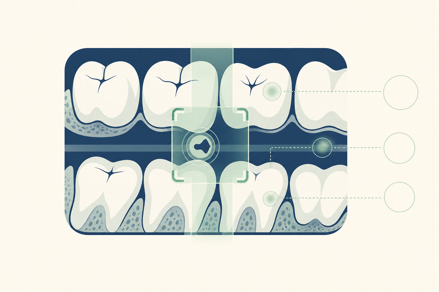
8-slide carousel · storyboard copy
AI on bitewing X-rays
Deep-learning models can identify patterns linked with caries on bitewing radiographs. Their value is greatest when they help a dentist notice and assess disease, not when they make an isolated treatment decision.
- AI is a reading aid, not a replacement for clinical examination.
- A bright mark on an X-ray is not automatically a cavity requiring a filling.
Why this matters
Bitewings show the crowns of the upper and lower premolars and molars, including the contact areas where tooth decay can hide. X-rays may reveal mineral loss in enamel or dentine before a lesion is obvious in the mouth.
- AI systems commonly use convolutional neural networks, or CNNs, to analyse pixel patterns.
- High AUC in a study describes separation between labelled cases. It does not prove that every patient benefits or that treatment decisions improve.
- The key comparison is stand-alone AI performance versus a dentist reading the image with AI assistance.
The clinical protocol
A safe reading combines the patient history, examination and radiograph rather than relying on one image label.
- The dentist reviews symptoms, diet, fluoride exposure, previous fillings, dry mouth risk and past caries.
- Bitewings are taken with the sensor positioned to show open contacts and the alveolar crest, while limiting radiation to what is clinically justified.
- The dentist checks enamel, the dentinoenamel junction and dentine on each proximal surface, then compares the finding with the visible tooth surface.
- If AI highlights a region, the dentist verifies image quality, considers overlapping anatomy and checks for restorations or artefacts.
- The diagnosis is recorded as sound, non-cavitated caries, cavitated caries, or another finding such as cervical burnout or a restoration margin.
The one rule that matters
Do not treat the software output. Treat the confirmed disease and its activity.
- A non-cavitated enamel lesion may be managed with plaque control, fluoride toothpaste, dietary changes and monitoring.
- A cavitated lesion, or decay extending into dentine with a risk of progression, may need operative treatment using a filling material such as resin composite or glass ionomer, depending on the tooth and site.
- A sound surface should not receive a filling simply because an algorithm marked it as suspicious.
Mythbuster: more sensitivity is always better
Not necessarily. Detecting more possible lesions can help prevent missed disease, but false-positive alerts can lead to extra radiographs, anxiety, unnecessary drilling and loss of healthy tooth structure.
- The dentist must balance sensitivity with specificity, image quality, lesion depth, cavitation and caries risk.
- AI can also reproduce errors from its training data, including differences in sensors, restorations, tooth shapes and patient populations.
- The most useful role is a second check that supports consistent review and discussion with the patient.
Make the result specific to you
Ask what the highlighted area represents in your tooth, not only whether the software says positive or negative.
- Is the lesion on the mesial or distal surface, and is it limited to enamel or extending into dentine?
- Can the surface be cleaned and monitored, or is there cavitation that prevents control?
- What preventive plan fits your risk, such as twice-daily fluoride toothpaste, reduced frequency of sugary drinks and snacks, and interdental cleaning?
- Use an interdental brush where the gap allows. Use floss where the contact is too tight for a brush. Spit after brushing and do not rinse.
When to see a dentist
Arrange a dental assessment when an X-ray shows a possible lesion, or sooner if you have toothache, sensitivity that lingers, food trapping, swelling, a broken tooth or a visible hole.
- The dentist may inspect the surface, dry it, use gentle visual and tactile examination, review earlier images and decide whether monitoring or treatment is appropriate.
- Urgent care is needed for facial swelling, fever, difficulty swallowing or difficulty breathing.
- If treatment is recommended, ask how the diagnosis was confirmed and what would happen if the area were monitored.
So, do the models help?
Yes, when they help dentists detect and document possible caries while the dentist remains responsible for diagnosis, risk assessment and treatment. They do not justify automatic fillings, and agreement with a dentist is not the same as clinical benefit.
- Bring the bitewing report and ask which tooth surface and tissue depth are involved.
- Use AI as an extra pair of eyes, with the final decision based on your mouth, your risk and the evidence on the image.
- Book a clinical review if you have symptoms or a new radiographic finding.