Composite or Porcelain Veneers
Composite veneers can be placed chairside in one visit and repaired directly. Porcelain veneers usually require preparation, laboratory fabrication and a second appointment, but offer greater colour stability and surface hardness. The right choice depends on enamel, bite, tooth position, disease control and the patient’s tolerance for maintenance, not on a universal ranking of materials.
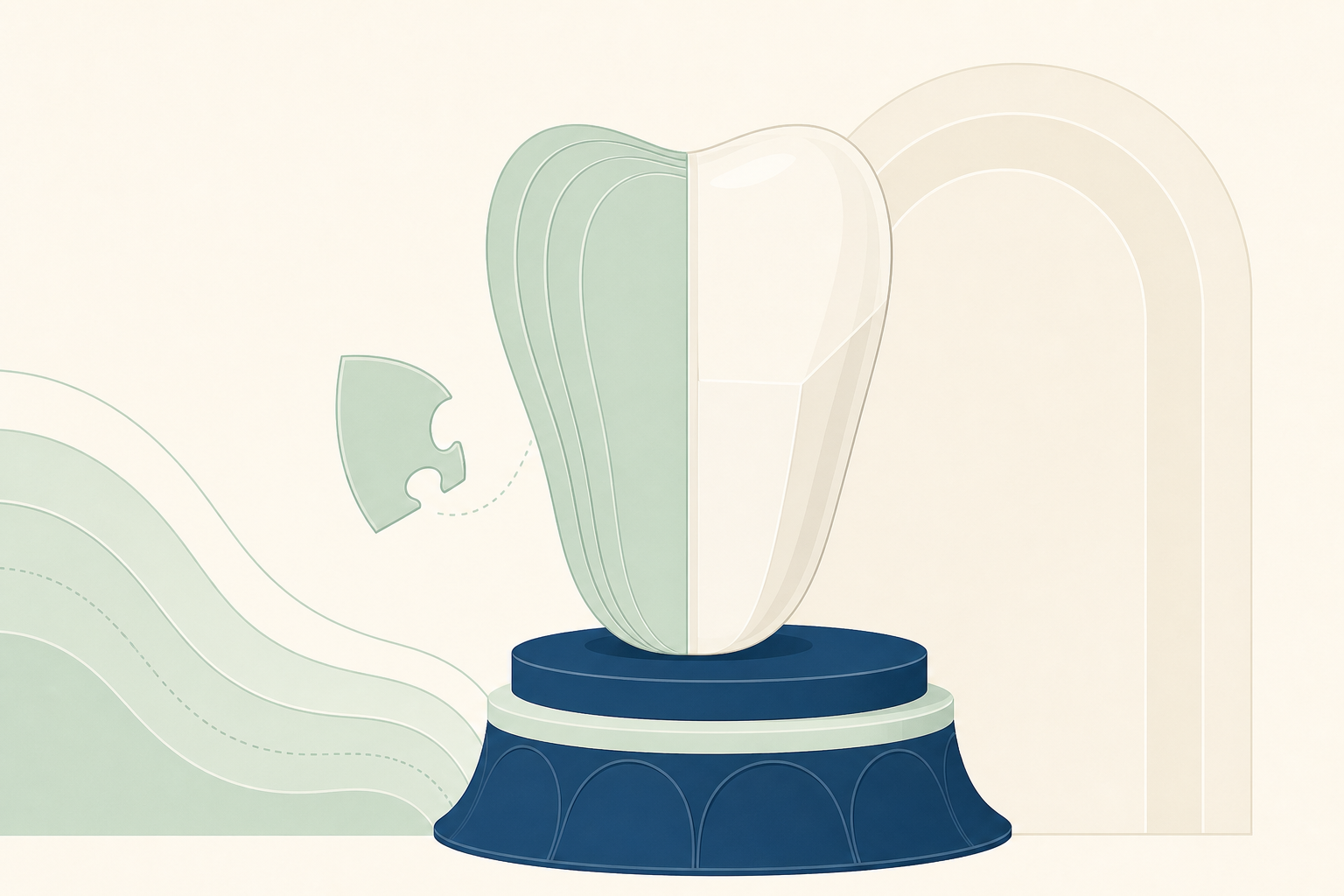
The decision starts with the tooth, not the material
A veneer is a bonded facial covering for the visible enamel of a tooth, most often an anterior incisor or canine. It can alter colour, contour, length, width and small spaces, but it does not correct the cause of every smile concern. The examination should identify caries, defective restorations, enamel thickness, cracks, erosion, abrasion, gingival position, tooth mobility and the relationship between the upper and lower incisors. The clinician also assesses parafunction, including clenching and grinding, because repeated loading can fracture either the veneer or the tooth beneath it. Active caries, untreated periodontal inflammation, poor plaque control and unstable occlusion should be managed before elective veneering. A rotated tooth, a severe deep bite, a large loss of tooth structure or a tooth with pulpal disease may require orthodontics, a restoration with greater coverage, endodontic treatment or no cosmetic treatment at all. Shade analysis should precede any whitening because composite and porcelain do not lighten after placement. The most conservative predictable result is usually achieved when the design remains within enamel and the facial surface is aligned with the patient’s existing occlusion.
The material is only one part of the decision. Tooth structure, disease control and loading determine whether a veneer is sensible.
Composite is a direct, repairable resin restoration
A direct composite veneer is built on the tooth from resin-matrix composite, usually containing inorganic glass or ceramic filler particles within a polymer resin. The tooth is isolated, cleaned and selectively prepared when needed. The enamel is etched with phosphoric acid, rinsed and dried according to the adhesive system, then treated with bonding resin. Composite is placed in increments or layers, shaped to reproduce the cervical contour, line angles, incisal translucency and contact point. Each increment is light-cured, then the restoration is finished and polished. The main clinical advantage is control at the chairside. The clinician can modify length, contour and shade during the appointment, and a chipped area can usually be roughened, etched, bonded and repaired with fresh composite. Little or no enamel may be removed in a suitable case, although adding material without adequate space can produce overcontour, plaque retention or an unstable bite. Composite is useful for a small enamel fracture, a localized discolouration, a modest diastema or a patient who needs a reversible trial of shape. Its limitations are water uptake, staining at the resin surface, wear, marginal breakdown and sensitivity to technique, especially when moisture contaminates the bonding field.
Composite turns the restoration into a chairside sculpture: fast to place, easy to revise, and dependent on meticulous bonding and polishing.
Porcelain is a laboratory-made bonded ceramic
A porcelain veneer is an indirect restoration fabricated from dental ceramic, commonly feldspathic porcelain or a glass ceramic such as lithium disilicate. The preparation is planned from a diagnostic wax-up or digital design so that reduction is controlled rather than guessed. Depending on the case, the facial enamel is reduced selectively, the incisal edge is preserved or modified, and the finish line is placed in a cleansable position. The prepared surfaces are recorded by an impression or intraoral scan, and a provisional restoration may be provided while the laboratory makes the veneer. At delivery, the clinician evaluates the fit, proximal contacts, marginal adaptation, shade, shape and occlusion before bonding. The internal ceramic surface is conditioned according to its composition, often by hydrofluoric acid etching followed by silane for glass ceramics. The enamel and dentine are treated with the selected adhesive protocol, and a light-cured or dual-cured resin cement is used where appropriate. Excess cement is removed before final curing and polishing. Ceramic offers a highly stable glazed or polished surface and can reproduce depth, opacity and translucency with precision. It is not indestructible. A thin ceramic edge can chip, and a debonded veneer may not be reusable.
Porcelain moves part of the work to the laboratory, where ceramic thickness, optical layering and surface texture can be controlled before delivery.
One visit versus two is a real clinical trade-off
The time difference is not simply convenience. Direct composite can often be completed in one appointment because the clinician creates and finishes the restoration in the mouth. That makes it appropriate when a patient needs a rapid change, when the design is limited, or when the final form is still being tested. The same chairside access also makes later additions and repairs straightforward, although a repair may remain visible if the surrounding composite has changed colour. Porcelain usually requires a preparation and records appointment followed by a separate fitting appointment. The laboratory stage permits controlled anatomy, contacts, translucency and symmetry across several teeth. It also introduces provisional wear, a second anaesthetic or isolation episode where required, and the risk that a provisional loosens or the final veneer needs adjustment. A laboratory does not remove the need for clinical judgement. If the preparation is too deep, the margin is poorly positioned, or the occlusion is not verified, a well-made ceramic veneer can still fail. For a patient attending from another GCC city or travelling for treatment, the appointments, provisional period and review visit should be treated as part of the treatment burden. The shortest schedule is not automatically the safest or most conservative schedule.
The chairside option saves laboratory time. The laboratory option spends more time before delivery to refine the final result.
Repairability and longevity pull in opposite directions
Composite generally has the stronger repair pathway. If an incisal corner fractures, the surface can be cleaned, roughened, conditioned and rebuilt with compatible resin. Small marginal defects may be polished or added to without replacing the entire veneer. This is valuable when the tooth is young, when the patient’s occlusion is changing, or when preserving options matters. Repair does not make composite maintenance-free. Repeated staining, loss of polish, wear at the incisal edge and recurrent fracture can require periodic refinishing or replacement. Porcelain is more resistant to surface staining and usually retains its texture and gloss well when properly finished. Its rigidity and bonded support can provide a durable restoration when enamel bonding, thickness, occlusion and patient selection are favourable. However, a fracture through the ceramic or a failure involving the bond often requires removal and remaking. Repair systems exist, but the colour match and strength may be less predictable than the original laboratory restoration. Neither material has a guaranteed service life. Longevity is affected by remaining enamel, adhesive isolation, ceramic design, composite handling, bruxism, diet, oral hygiene, trauma and review. A night guard may be indicated for a patient who clenches or grinds, but it does not make an overloaded design safe.
Composite usually fails in a more repairable way. Porcelain usually offers better surface stability, but a major fracture is less forgiving.
Who tends to suit each option
Composite is often suited to a patient with a limited number of teeth to change, a small fracture or space, adequate enamel and a low to moderate functional demand. It can be a rational first treatment when the patient wants to test a new contour, expects future changes, or values direct repair. It is also useful when the clinician can preserve enamel and establish a stable emergence profile without creating a bulky facial surface. Porcelain is often suited to a patient with multiple stable aesthetic concerns, such as persistent intrinsic discoloration, several worn or undersized anterior teeth, or a need for closely matched shape and optical behaviour across a smile. It may be preferred where stain resistance and long-term surface polish are high priorities and where sufficient enamel remains for reliable adhesive bonding. The patient must accept preparation, laboratory fabrication and the possibility that replacement will require further treatment. A patient with untreated bruxism, active erosion from gastric reflux, frequent acidic exposure, poor plaque control or an unstable bite may suit neither option yet. The sequence may need to begin with disease control, occlusal assessment, dietary change, orthodontics, whitening, remineralisation support or treatment of the underlying medical problem.
Composite suits controlled change and repair. Porcelain suits a stable, carefully planned result when the patient accepts an irreversible laboratory process.
Upkeep is clinical maintenance, not a cosmetic afterthought
Veneered teeth still have a gingival margin, proximal surfaces and often exposed enamel or dentine. Plaque can accumulate where the restoration is overcontoured or where the finishing line is rough. Brush twice daily with fluoride toothpaste, then spit rather than rinse so fluoride remains available at the tooth surface. Use interdental brushes where the gap allows, because they clean a broader contact area; floss remains appropriate where an interdental brush cannot pass or where the contact is tight. Do not snap floss through a contact or pull it upward against a veneer margin. A soft or medium toothbrush, non-abrasive toothpaste and regular professional review help preserve the polish and monitor the gingivae, margins, contacts and opposing teeth. Avoid using the veneered teeth as tools for cutting packaging or biting hard objects. Coffee, tea, tobacco and strongly coloured foods can stain exposed composite and the cement or margins, although porcelain itself is comparatively colour stable. Whitening after placement may lighten natural teeth without changing the veneer shade, creating mismatch. Review is also an opportunity to check occlusion and signs of parafunction. A night guard may protect a selected patient, but it cannot compensate for poor diagnosis, inadequate material thickness or a destructive bite.
The restoration ends at the appointment. The treatment continues through plaque control, sensible loading and review of the margins.
The answer is a material choice with boundaries
Choose direct composite when the clinical problem is limited, enamel can be preserved, chairside modification is valuable and repairability matters more than maximum surface stability. Choose porcelain when the aesthetic plan is broader, the tooth and bite are stable, enamel bonding is available and the patient accepts a laboratory process with less forgiving repair. In both cases, the design should respect the cervical anatomy, the gingival architecture, the incisal edge and the path of the opposing teeth. Neither veneer should be used to conceal untreated disease, replace orthodontic movement that is needed for function, or cover a tooth that requires a crown, endodontic treatment or periodontal care. Very dark substrates, minimal enamel, severe crowding, extensive structural loss and active parafunction can make predictable bonding difficult. In those situations, doing less may protect more tooth tissue. For clinicians and informed patients in the UAE and GCC, the practical conclusion is clear: compare the biological cost, appointment burden, repair pathway and maintenance demands before comparing the smile photographs. A sound decision is the one that solves the defined problem while leaving the greatest number of future options open.
The best veneer is not the most expensive-looking one. It is the least invasive restoration that remains stable, cleansable, repairable and appropriate to the bite.
Key findings
- Composite veneers are direct resin restorations that can often be completed in one visit and repaired chairside.
- Porcelain veneers are laboratory-made ceramic restorations that usually require preparation, provisionalisation and a second appointment.
- Porcelain is generally more resistant to surface staining and loss of polish, while composite usually offers easier direct repair and modification.
- Both options depend on enamel preservation, moisture control, accurate contour, stable occlusion and control of caries, periodontal inflammation and parafunction.
- Neither option is appropriate as a substitute for treating active disease, severe malposition, major structural loss or an unstable bite.