Catching Decay Years Earlier: The Quiet Revolution in Dental Scanning
New imaging can spot trouble long before it hurts or shows on an X-ray. This is one to file firmly under "closer than you think" and ask your dentist about.
8-slide carousel · storyboard copy
Decay can start quietly
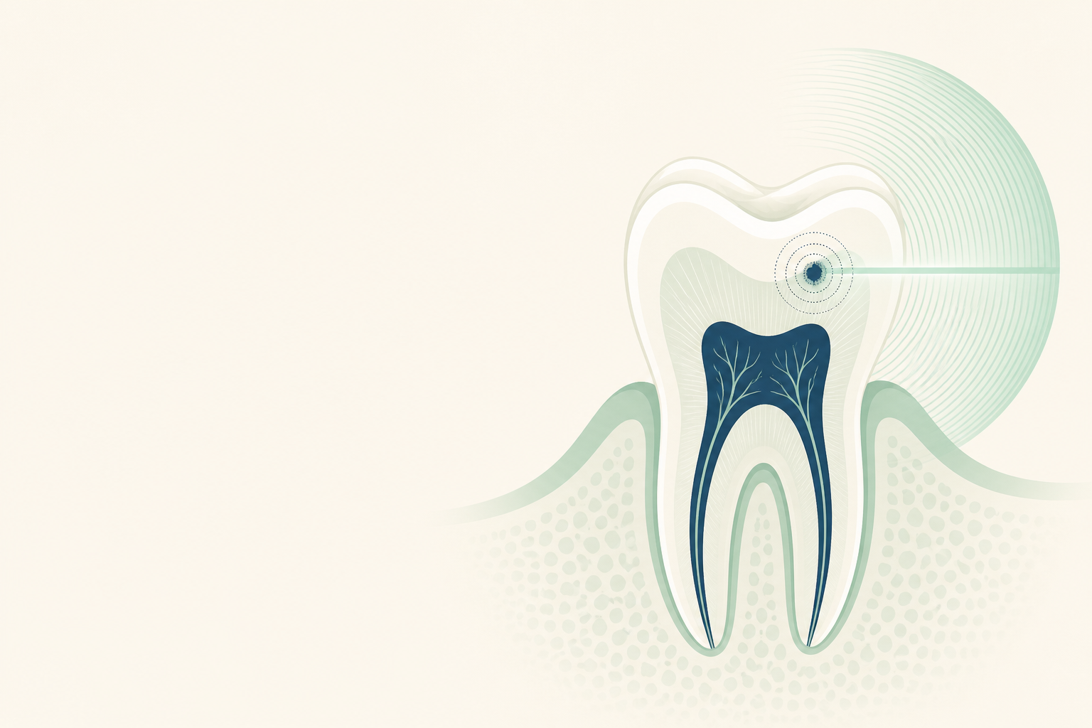
A tooth may look normal, feel normal and still be losing mineral beneath its surface. Near-infrared transillumination uses light to reveal some of these early changes before a cavity becomes obvious.
- The target is demineralised enamel and dentine.
- The scan uses non-ionising near-infrared light.
- It is an aid to examination, not a standalone diagnosis.
Why it matters before pain
Tooth decay begins when acids from plaque bacteria dissolve hydroxyapatite, the calcium-phosphate mineral in enamel. Early lesions may be limited to enamel and can sometimes be stabilised without drilling. Once decay reaches the enamel-dentine junction or dentine, progression and restorative treatment become more likely.
- Common hidden sites include the contact areas between premolars and molars.
- Pain usually indicates deeper irritation, not the beginning of decay.
- Bitewing radiographs remain important, especially for proximal lesions.
The protocol in steps
The dentist first examines the teeth, cleans and dries the surfaces, and records risk factors such as frequent sugar exposure, low fluoride exposure, plaque accumulation, dry mouth, previous decay and existing fillings. The scanner is then positioned against or over the tooth.
- Near-infrared light passes through enamel and dentine.
- Sound and demineralised tissues scatter and absorb the light differently.
- An infrared-sensitive camera records a contrasting image.
- The dentist compares the image with the clinical examination and, when indicated, bitewing radiographs.
One rule matters most
A dark or bright area on a scan is a finding, not automatically a cavity requiring a filling. The dentist must judge whether the lesion is active, how deep it is, whether the surface is intact, and whether it can be monitored or treated conservatively.
- Early enamel change may be managed with plaque control, dietary change and fluoride toothpaste.
- Selected non-cavitated lesions may be considered for resin infiltration.
- A cavitated lesion that cannot be cleaned may need a restoration.
- The same area can be rescanned over time to assess change.
Mythbuster: it replaces X-rays
It does not. Near-infrared imaging can improve visualisation of some proximal and occlusal lesions, particularly early enamel changes, but it has limits. Staining, restorations, tooth anatomy and lesion depth can affect interpretation. Radiographs and clinical examination still have roles.
- The scan does not use X-rays.
- It cannot reliably identify every lesion.
- A bitewing radiograph may be needed when dentine involvement or hidden proximal decay is suspected.
- No imaging method removes the need for a dentist’s diagnosis.
Make the finding useful
Ask which tooth surface is affected, whether the enamel surface is cavitated, whether the lesion reaches dentine, and when it should be reviewed. At home, brush twice daily with fluoride toothpaste, spit rather than rinse, and clean between teeth daily.
- Use an interdental brush where the space allows.
- Use floss where contacts are too tight for an interdental brush.
- Reduce how often teeth are exposed to sugary foods and drinks.
- Do not treat hydroxyapatite as equivalent to fluoride.
When to see a dentist
Book an examination if you have sensitivity, food trapping, a rough or broken area, a visible white or brown change, persistent bad breath, or a history of repeated decay. Do not wait for pain. At the visit, the dentist can decide whether near-infrared imaging, bitewing radiographs or another test is appropriate.
- Tell the dentist about dry mouth, reflux, orthodontic appliances and frequent snacking.
- Existing crowns, fillings and sealants may change how images are read.
- Urgent care is appropriate for facial swelling, fever, or difficulty swallowing or breathing.
Closer than you think
The quiet revolution is not a magic scan. It is earlier information about mineral loss, before a small lesion becomes a larger repair. If your clinic offers near-infrared dental imaging, ask what it shows, what it cannot show, and how the result changes your treatment plan.
- Early detection can support prevention and minimally invasive care.
- Your dentist should interpret the image alongside examination and risk assessment.
- Save the question for your next dental check-up: "Can you monitor early changes before they become cavities?"